


Research Article | Avian Medicine | Veterinary Pathology
Authors: Pouneh Hajipour, Yousef Toutounkar, Navid Khosroabadi, Omid Dezfoulian, and Jamshid Razmyar
Journal: Case Reports in Veterinary Medicine
Publication Year: 2026
DOI: https://doi.org/10.1155/crve/5662048
Article Type: Open Access Case Report
Hepatic lymphoma is an uncommon neoplastic condition in birds, and its diagnosis can be particularly challenging in mynahs, in which marked liver enlargement may be associated with several metabolic and neoplastic disorders. This case report describes hepatic B-cell lymphoma in a 14-year-old common mynah (Acridotheres tristis) and highlights the importance of a comprehensive diagnostic approach to avian hepatic masses.
The case was investigated using clinical examination, diagnostic imaging, gross and histopathological evaluation, immunohistochemistry, and molecular testing for selected avian oncogenic viruses.
A 14-year-old male common mynah was presented with lethargy, altered vocalization, poor body condition, coelomic distension associated with fluid accumulation, respiratory distress, chronic ataxia, and left-leg lameness.
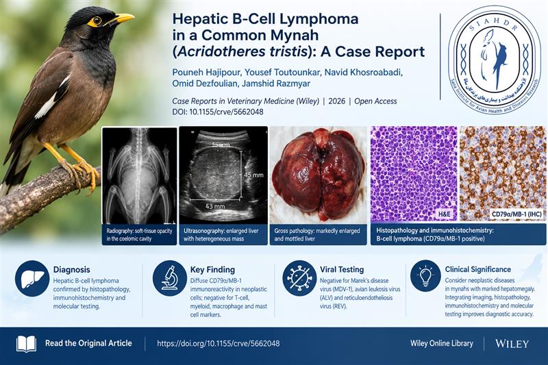
Radiographic examination revealed a large soft-tissue opacity within the coelomic cavity, displacing the intestinal loops and compressing the thoracoabdominal air sacs. Ultrasonography subsequently identified a markedly enlarged liver containing a heterogeneous mass measuring approximately 53 × 45 mm and occupying a substantial portion of the coelomic cavity.
Because of the severity of the lesion and the bird’s poor clinical condition, euthanasia was performed at the owner's request. Necropsy revealed severe hepatomegaly, with the liver extensively occupying the coelomic cavity.
Histopathological examination demonstrated extensive replacement of the hepatic parenchyma by densely packed neoplastic mononuclear cells, accompanied by disruption of the normal hepatic architecture and hepatocellular necrosis.
Immunohistochemical evaluation showed diffuse cytoplasmic immunoreactivity for CD79α/MB-1 in the neoplastic cells. The tumor cells did not demonstrate evidence of T-cell differentiation, and the overall immunohistochemical and histochemical profile did not support macrophage, myeloid, or mast-cell origin. These findings supported the diagnosis of hepatic B-cell lymphoma.
Molecular testing of liver tissue was negative for Marek’s disease virus type 1 (MDV-1), avian leukosis virus (ALV), and reticuloendotheliosis virus (REV). No neoplastic infiltration was identified in the examined spleen, kidneys, intestines, lungs, or heart; therefore, the liver was the only confirmed site of lymphoma among the tissues examined.
This report emphasizes that severe hepatomegaly in mynahs should not automatically be attributed to iron storage disease or other metabolic hepatopathies. Neoplastic conditions, including lymphoma, should also be considered in the differential diagnosis.
The findings demonstrate the complementary value of diagnostic imaging, histopathology, immunohistochemistry, and targeted molecular testing in characterizing hepatic masses in birds. A tissue-based diagnostic approach is particularly important when clinical signs and imaging findings alone cannot reliably establish the nature or lineage of a lesion.
To the authors’ knowledge, this is the first reported case of hepatic B-cell lymphoma in a common mynah supported by immunohistochemical characterization.
Hepatic B-cell lymphoma should be considered among the differential diagnoses for marked hepatomegaly and hepatic masses in common mynahs. Integrating imaging findings with histopathological and immunohistochemical evidence can improve diagnostic accuracy and help distinguish lymphoma from other hepatic disorders in avian patients.
Avian oncology; Common mynah; Acridotheres tristis; B-cell lymphoma; Hepatomegaly; Immunohistochemistry; Histopathology; Companion birds.
For the complete methodology, diagnostic images, histopathological findings, and references, please consult the original open-access publication:
Read the full article: https://doi.org/10.1155/crve/5662048
Institutional affiliation: Sana Institute for Avian Health and Diseases Research, Tehran, Iran.